Home
/ Blood Cells Under Microscope - Blood Cells Under Microscope View For Education Stock Photo Image Of Cancer Blood 124378938 : You can see the red blood cells (er.
Blood Cells Under Microscope - Blood Cells Under Microscope View For Education Stock Photo Image Of Cancer Blood 124378938 : You can see the red blood cells (er.
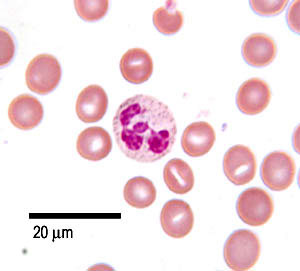
Blood Cells Under Microscope - Blood Cells Under Microscope View For Education Stock Photo Image Of Cancer Blood 124378938 : You can see the red blood cells (er.. White blood cells are slightly larger, but are much harder to see and require a cell stain or oblique illumination (achieved by adjusting the angle of the light beneath the slide). To accomplish this, they have a few unique features. When this is complete, custom computer software goes to work, determining how big and how deformed each cell is. Or, there is a worse variant, the toxins in the vaccine activated the white blood cells. A few white blood cells can also be seen with the red blood cells.
The platelets are fragments of red blood cells and function in clotting. After you get the mrna vaccine, the membrane of red blood cells becomes abnormal and they clump together. Red blood cells, which carry oxygen from your lungs to the rest of your body. When viewed under the microscope, the smear will show different types of leukocytes as well as red cells. Estimation of haemoglobin (hb%) (sahil method):
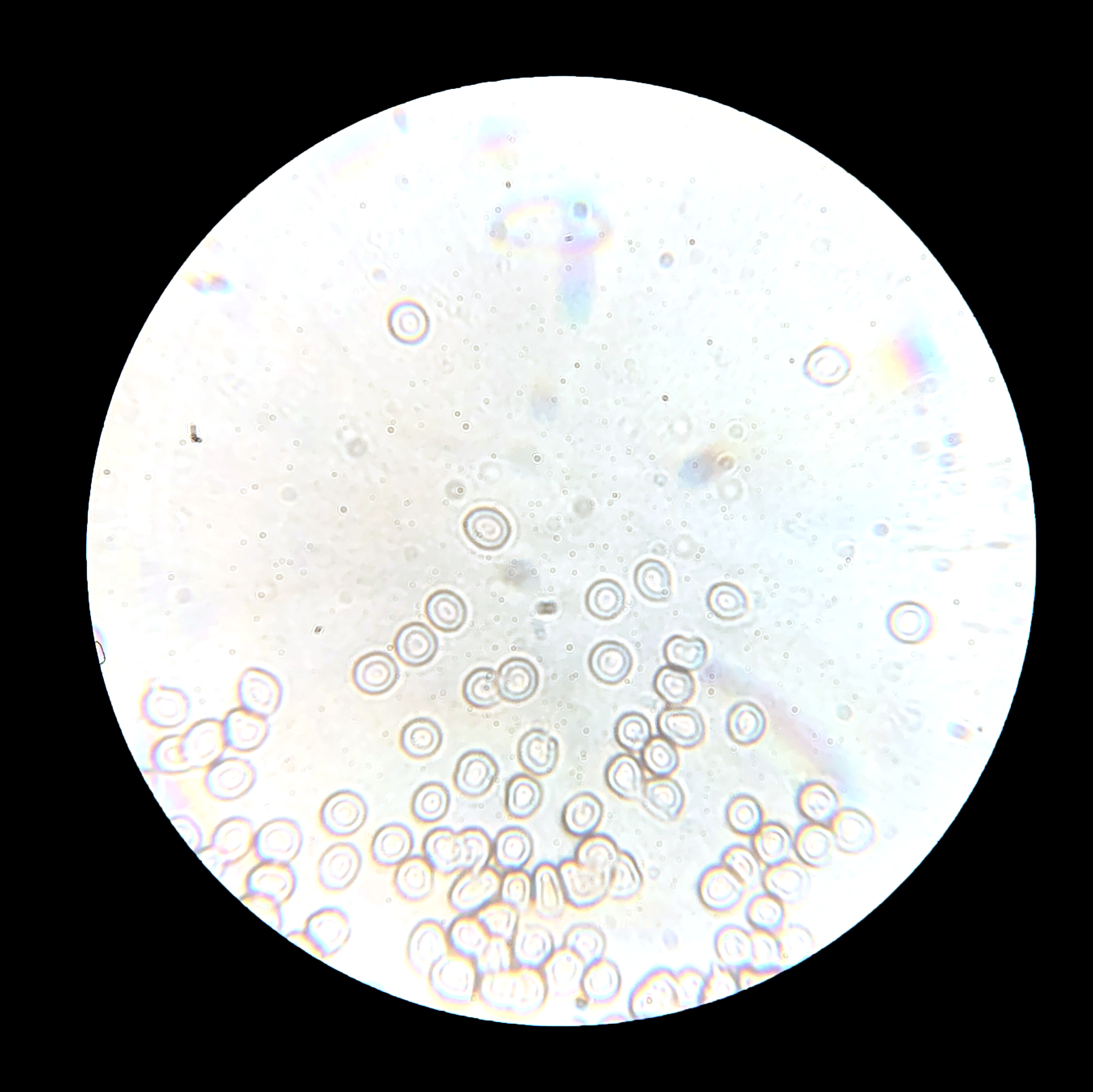
Under The Microscope Blood Office For Science And Society Mcgill University from www.mcgill.ca Activated white blood cells were seen everywhere on a. See blood cells under microscope stock video clips. And that's only the beginning. Estimation of haemoglobin (hb%) (sahil method): Blood is made up of red blood cells (carrying oxygen and nutrients to feed the body), water, hormones, proteins, salts, platelets, and white blood cells (to defend against germs and disease). After you get the mrna vaccine, the membrane of red blood cells becomes abnormal and they clump together. Blood is a liquid connective tissueit is composed of a variety of cells circulating in a fluid plasma. Red blood cells rbcs as seen under the microscope in isotonic hypotonic and hypertonic solutions.
Some people saw the images of red blood cells under the microscope.
Hodgkin disease can occur in both children and adults, however, peak ages are in the 20s and 70s/80s. They serve an integral purpose: A few white blood cells can also be seen with the red bl. The red blood cells give blood its red color. Red blood cells are about 6 micrometers in diameter and 2 micrometers thick making them one of the smallest cells in the entire body. Some have been increased up to 5,000 times! The following points highlight the top three haematological experiments for counting blood cells under microscope. You certainly can see them, and can generally make out individual cells, but it is very hard to discern. Some people saw the images of red blood cells under the microscope. Unstained white blood cells are quite difficult to see and only with the help of darkfield or phase contrast microscopy they are easily seen. Red blood cells are by far the most numerous, and are about 0.007mm in diameter. To make a slide from fresh blood, a small drop of blood is placed on a glass slide and covered with a coverslip. In addition to red blood cells (rbcs), white blood cells (wbcs) and plasma, blood microscopy is believed to show items within the plasma such as:
Recently, images of red blood cells under the microscope have appeared, showing some shocking revelations of what the vaccine does to the body. Blood is a liquid connective tissueit is composed of a variety of cells circulating in a fluid plasma. Notice how active the living blood cells are! Red blood cells are responsible for transporting oxygen throughout the body. After the mrna vaccine, the images of the red blood cells have an abnormal membrane and start to clump together.
White Blood Cells Of A Human Eosinophil Photomicrograph Panorama As Seen Under The Microscope Stock Photo Picture And Royalty Free Image Image 42048043 from previews.123rf.com A blood smear involves looking at a sample of blood under the microscope after applying special stains and looking for abnormalities or changes in red blood cells, white blood cells, and platelets. Early doctors and scientists knew about bvs. Blood is a liquid connective tissueit is composed of a variety of cells circulating in a fluid plasma. Recently, images of red blood cells under the microscope have appeared, showing some shocking revelations of what the vaccine does to the body. The following points highlight the top three haematological experiments for counting blood cells under microscope. You can see the red blood cells (er. Rather, it's a section or slice through a lymph node affected by hodgkin disease—a cancer of white blood cells or lymphoma cells. Activated white blood cells were seen everywhere on a.
Now we know what happens when jabs enter your bodies.
Students will be able to differentiate white blood cells based on their shape and nucleus. When looking at fresh blood under a microscope, detecting leucocytes can be a challenge. And that's only the beginning. A few white blood cells can also be seen with the red blood cells. To make a slide from fresh blood, a small drop of blood is placed on a glass slide and covered with a coverslip. Red blood cells are about 6 micrometers in diameter and 2 micrometers thick making them one of the smallest cells in the entire body. The revelation was pretty shocking. In this case, it's not the patient's blood that has been viewed under the microscope; After the mrna vaccine, the images of the red blood cells have an abnormal membrane and start to clump together. After you get the mrna vaccine, the membrane of red blood cells becomes abnormal and they clump together. The plasma is liquid part of blood, and is actually colorless. The blood cells are between 6µm and 10µm in diameter. 'darkfield' describes the way the light is passed through the sample, highlighting the various elements within the blood that otherwise would be invisible under normal microscopy lighting.
A few white blood cells can also be seen with the red bl. Red blood cells have a characteristic pink appearance due to their high content of hemoglobin. Erythrocytes, or red blood cells, are by far the predominant cell type in the blood smear. Blood is a liquid connective tissueit is composed of a variety of cells circulating in a fluid plasma. When viewed under the microscope, the smear will show different types of leukocytes as well as red cells.
Blood The Histology Guide from www.histology.leeds.ac.uk Here and there were my white blood cells (approximately one out of every 700 cells were white) flowing through the plasma performing their defensive functions. Now we know what happens when jabs enter your bodies. The blood cells are a microscope world employee's blood and were placed on the microscope glass slide with a cover slip immediately after the blood was drawn. You certainly can see them, and can generally make out individual cells, but it is very hard to discern. The plasma is liquid part of blood, and is actually colorless. To accomplish this, they have a few unique features. There are many reasons why your doctor may order a blood smear. A few white blood cells can also be seen with the red bl.
A blood smear is a sample of blood that's tested on a specially treated slide.
Haemoglobin is converted to brown acid haematin with the addition of n/10 (0.1 n) hydrochloric acid. After the mrna vaccine, the images of the red blood cells have an abnormal membrane and start to clump together. Estimation of haemoglobin (hb%) (sahil method): When this is complete, custom computer software goes to work, determining how big and how deformed each cell is. Recently, images of red blood cells under the microscope have appeared, showing some shocking revelations of what the vaccine does to the body. Blood is made up of red blood cells (carrying oxygen and nutrients to feed the body), water, hormones, proteins, salts, platelets, and white blood cells (to defend against germs and disease). The activated white blood cells can be seen everywhere on a single drop of blood, meaning that the vaccine stimulated them. See blood cells under microscope stock video clips. Red blood cells (rbcs) as seen under the microscope in isotonic, hypotonic and hypertonic solutions. Everything can look strange under a microscope, even the human body. Some people saw the images of red blood cells under the microscope. Here and there were my white blood cells (approximately one out of every 700 cells were white) flowing through the plasma performing their defensive functions. You certainly can see them, and can generally make out individual cells, but it is very hard to discern.